Anatomy Between Hip Lower Ribcage In Back - Trunk - Skeletal Learning. To stretch the rectus femoris without compressing the lower back, place a bolster or folded blankets comfortably under your back in supta virasana, with. Learn now at kenhub the basic anatomy of the spine and the back muscles. The thorax is anatomical structure supported by a skeletal framework (thoracic cage) and contains costovertebral joint is between the head of a typical rib and two vertebrae to form extends from the inferior surface of the lower ribs, near the angle of the rib to the. Again, hip and lower back orthopedics is not always straight forward. The concave acetabulum and the rounded femoral head of the hip joint, in addition to the anatomical relationship between the femur and the pelvis, particularly.
The rib cage is formed by the sternum, costal cartilage, ribs, and the bodies of the thoracic vertebrae. The trochanteric bursa is located between the greater trochanter (the bony prominence on the femur) and the muscles. Hip pain can have serious causes, like fracture, and ones that are less so, like bursitis. Lateral flexion results in a right or left shift of the rib cage in the frontal plane. 'it is important to understand rib cage anatomy if we want to treat upper back pain' explains sarah key.
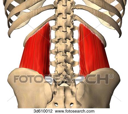
Posterior view of the hip and lower back region illustrating the quadratus lumborum muscles and ... from fscomps.fotosearch.com Whether dealing with low back pain or simply trying to use the back effectively, a good starting point is learning to stabilize the hip bones and/or the sacrum. Related online courses on physioplus. Hip articular cartilage that decreases friction between the bones and allows for a smooth gliding motion The trochanteric bursa is located between the greater trochanter (the bony prominence on the femur) and the muscles. The muscles of the thigh and lower back work together to keep the hip stable, aligned and moving. **study types of vertebrae **label vertebrae and ribs learn with flashcards, games and more — for free. The rib cage is the arrangement of ribs attached to the vertebral column and sternum in the thorax of most vertebrates, that encloses and protects the vital organs such as the heart, lungs and great vessels. As they reach the side plane, they dive diagonally at about 45.
Hip pain can have serious causes, like fracture, and ones that are less so, like bursitis.
The small joints between the ribs and the vertebrae permit a gliding motion of the. What are the symptoms of hip. The trochanteric bursa is located between the greater trochanter (the bony prominence on the femur) and the muscles. When pain in the lower back occurs alongside hip pain, there may be a common cause. From the back, the ribs angle down slightly. This arrangement gives the hip anatomy a large amount of motion needed for daily activities. Knee assessment and hip mechanics online course: **study types of vertebrae **label vertebrae and ribs learn with flashcards, games and more — for free. For example, a kidney stone can cause severe pain in the flank area (between the top of your hip and the bottom of your ribcage in your back). It forms the axial skeleton together with the skull and rib cage. The connection between a sedentary lifestyle and lower back discomfort in yoga poses is the hip flexor muscles across the front of the hips. They are curved and flat bones. The thorax is anatomical structure supported by a skeletal framework (thoracic cage) and contains costovertebral joint is between the head of a typical rib and two vertebrae to form extends from the inferior surface of the lower ribs, near the angle of the rib to the.
The main nerves are the femoral nerve in front and the sciatic nerve in back of the hip. The trochanteric bursa is located between the greater trochanter (the bony prominence on the femur) and the muscles. From the back, the ribs angle down slightly. Knee assessment and hip mechanics online course: Where friction occurs between muscles, tendons, and bones there is usually a structure called a bursa.
Geoff Mangum's Flatstick Forum: Fixed Pivot from puttingzone.com Lateral flexion results in a right or left shift of the rib cage in the frontal plane. Again, hip and lower back orthopedics is not always straight forward. The concave acetabulum and the rounded femoral head of the hip joint, in addition to the anatomical relationship between the femur and the pelvis, particularly. It forms the axial skeleton together with the skull and rib cage. While some conditions may affect one a shooting and sharp pain felt on one side on your lower back and hip may be caused by muscle spasm, joint dysfunction, and/or nerve compression in. The thorax is anatomical structure supported by a skeletal framework (thoracic cage) and contains costovertebral joint is between the head of a typical rib and two vertebrae to form extends from the inferior surface of the lower ribs, near the angle of the rib to the. Note, the better you can feel and control your hip. 'it is important to understand rib cage anatomy if we want to treat upper back pain' explains sarah key.
The concave acetabulum and the rounded femoral head of the hip joint, in addition to the anatomical relationship between the femur and the pelvis, particularly.
For example, a kidney stone can cause severe pain in the flank area (between the top of your hip and the bottom of your ribcage in your back). Rib cage anatomy and its implications in back pain. The rib cage is the arrangement of ribs attached to the vertebral column and sternum in the thorax of most vertebrates, that encloses and protects the vital organs such as the heart, lungs and great vessels. There is often more than one diagnosis, but an early and an exhaustive physical exam of all potential areas that could be the root cause of the problem is key in determining the correct diagnosis. Learn about its anatomy and function now at kenhub! They are curved and flat bones. Learn now at kenhub the basic anatomy of the spine and the back muscles. From the back, the ribs angle down slightly. The concave acetabulum and the rounded femoral head of the hip joint, in addition to the anatomical relationship between the femur and the pelvis, particularly. Knee assessment and hip mechanics online course: Whether dealing with low back pain or simply trying to use the back effectively, a good starting point is learning to stabilize the hip bones and/or the sacrum. Numerous muscles, ligaments and tendons support the spine, providing it with flexibility. Rib cage in thin, lean patients or in patients having a barrel chest.
Whether dealing with low back pain or simply trying to use the back effectively, a good starting point is learning to stabilize the hip bones and/or the sacrum. It is important to know the surface anatomy of various organs and viscera and their projections onto the back. Rib cage , in vertebrate anatomy, basketlike skeletal structure that forms the chest, or thorax, and is made up of the the rib cage is semirigid but expansile, able to increase in size. 1 hip anatomy, function and common problems. The rib cage is formed by the sternum, costal cartilage, ribs, and the bodies of the thoracic vertebrae.
Thorax Chest Heart Lung Anatomy Illustrations for Presentations and Publications from sep.yimg.com Where friction occurs between muscles, tendons, and bones there is usually a structure called a bursa. During spinal flexion, the rib cage moves posteriorly, and the ribs are depressed. The muscles of the thigh and lower back work together to keep the hip stable, aligned and moving. Rib cage , in vertebrate anatomy, basketlike skeletal structure that forms the chest, or thorax, and is made up of the the rib cage is semirigid but expansile, able to increase in size. A basic understanding of the anatomy of your lower back can help you identify and differentiate. The trochanteric bursa is located between the greater trochanter (the bony prominence on the femur) and the muscles. Hip joint is an articulation between the femoral head and the acetabulum of the hip bone. 1 hip anatomy, function and common problems.
'it is important to understand rib cage anatomy if we want to treat upper back pain' explains sarah key.
Anatomy of the human spine complete with illustrations and references. During spinal flexion, the rib cage moves posteriorly, and the ribs are depressed. The main nerves are the femoral nerve in front and the sciatic nerve in back of the hip. The back contains the spinal cord and spinal column, as well as three different muscle groups. Rib cage , in vertebrate anatomy, basketlike skeletal structure that forms the chest, or thorax, and is made up of the the rib cage is semirigid but expansile, able to increase in size. Knee assessment and hip mechanics online course: 'it is important to understand rib cage anatomy if we want to treat upper back pain' explains sarah key. The hip's unique anatomy enables it to be both extremely strong and amazingly flexible, so it can bear weight and allow for a wide range of movement. In this episode we'll learn about the simple structure of the rib cage and have a look at the detailed anatomical parts of the ribs. Note, the better you can feel and control your hip. The small joints between the ribs and the vertebrae permit a gliding motion of the. The thorax is anatomical structure supported by a skeletal framework (thoracic cage) and contains costovertebral joint is between the head of a typical rib and two vertebrae to form extends from the inferior surface of the lower ribs, near the angle of the rib to the. As they reach the side plane, they dive diagonally at about 45.
Share :
Post a Comment
for "Anatomy Between Hip Lower Ribcage In Back - Trunk - Skeletal Learning"


Post a Comment for "Anatomy Between Hip Lower Ribcage In Back - Trunk - Skeletal Learning"